Neuroconductor
Neuroconductor was developed in the Department of Biostatistics at Johns Hopkins University by John Muschelli (lead R project expert), Adrian Gherman (lead software engineer), Ciprian Crainiceanu and Brian Caffo (Principal Investigators). Neuroconductor (https://neuroconductor.org/) is an open-source platform for rapid testing and dissemination of reproducible computational imaging software. The goals of the project are to:
- provide a centralized repository of R software dedicated to image analysis;
- disseminate quickly software updates;
- educate a large, diverse community of scientists using detailed tutorials and short courses;
- ensure quality via automatic and manual quality controls; and
- promote reproducibility of image data analysis.
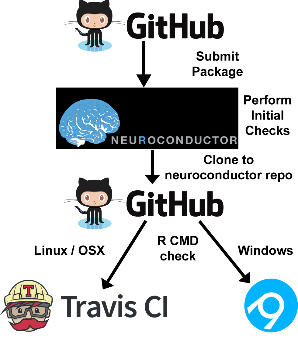
Based on the programming language R, Neuroconductor starts with 40 inter-operable packages that cover multiple areas of imaging including visualization, data processing and storage, and statistical inference. Neuroconductor accepts new R package submissions, which are subject to a formal review and continuous automated testing. Neuroconductor also provides data packages and packages designed to download data from online imaging repositories such as the Human Connectome Project (Along with all the other capabilities of R, Neuroconductor can be integrated with Shiny applications, reproducible reports, and journal articles. Neuroconductor is heavily influenced by the Bioconductor platform, though Neuroconductor was built later and takes advantage of new tools that were not available when Bioconductor was started. More precisely, the framework builds upon open source projects using git for version control, GitHub for package distribution, the devtools R package for package installation from GitHub, and Travis CI and Appveyor continuous integration services for checking packages and building binary packages for distribution on GitHub. The image below provides an overall view of the Neuroconductor structure.
Currently, there are tutorials for structural brain image analysis and the goal is to expand these tutorials into different imaging areas, with focus on functional magnetic resonance imaging (fMRI) and structural imaging of areas of the body other than the brain.

